Mending Broken Hearts with Cells and Bioprinting Technology
Good afternoon, everybody. Welcome to today's UTS Science in Focus, which is a free public lecture series showcasing the latest research from prominent UTS scientists and researchers. I'm Professor Joanne Tipper, the head of School of Biomedical Engineering and also the acting Dean of the Graduate Research School. And I'll be comparing today's event, collating the questions and asking our speakers the questions at the end of the talk. Today's topic is important for understanding and developing new technologies to address health care problems in the ageing population. Before I introduce our speakers today, I would like to acknowledge the Gadigal people of the Eora nation whose, upon whose ancestral lands our city campus at UTS now stands. I'd like to pay my respects to the elders, both past, present and also emerging, acknowledging them as the traditional owners, the custodians of knowledge for this land. Just a little bit of housekeeping before we begin today's session. It is an online only event, unfortunately. So do bear with us. If we have any technical issues, we'll work as quickly as we can to resolve them. And if you find you're not able to access the talk at any point, the easiest way to rectify that is to log out and then log back in again. That usually resolves these sorts of issues. We will be having a Q&A session at the end of the, the talks for about half an hour, if you would like to ask a question, then please type your question into the Q&A box on your Zoom control panel, and we'll do our best to answer all the questions at the end.
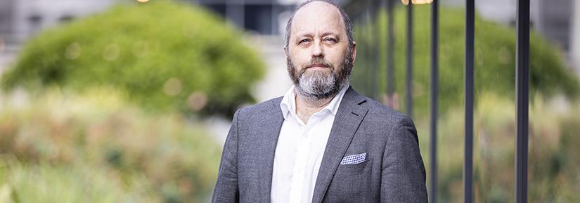
If you like somebody else's question, then you can upvote that question to make sure that it gets asked and you hear the answer. And you can do that by clicking on the little thumbs up symbol next to the question itself. And that will make that question rise up the the list. We will be recording the session today. However, we won't be recording any video or audio input from the audience. If you have any concerns about the recording of the event today, you can contact UTS at science.future@uts.edu.au. And the address is there on this line, so tell us any concerns that you have. So onto the interesting bit today, so our talk today is entitled Mending Broken Hearts with Cells and 3D Bioprinting Technology. Our speakers are Dr. Carmine Gentile from the School of Biomedical Engineering and then two artists who Carmine currently collaborates with, Paul Brown and Linda Dement. So I'm just going to give you a little bit of information about the speakers. So Carmine is actually a pharmacist and now a lecturer in the School of Biomedical Engineering at UTS. He leads the Cardiovascular Regeneration Group at UTS and also at the University of Sydney. He received his Bachelor's and Master's degrees from the University of Pisa in Italy and then his Ph.D. from the Medical University of South Carolina in the US. Since 2013, Carmine has worked in Australia, first at the Heart Research Institute, then at the University of Sydney and now at UTS. He's been supported by numerous awards and grants, working with a multidisciplinary team of scientists, industry partners and clinicians to quickly translate his research findings from bench to bedside.
He's an internationally recognised researcher, expert in the field of 3D printing and stem cell technologies, and his more recent studies focus on novel, molecular and cellular approaches to treat cardiovascular disease, including myocardial infarction and heart failure. These studies are based on the use of mini hearts, which he developed as bio-inks for human heart tissues. And that's the subject of today's presentation. Paul Brown is an Earth scientist, a creative artist and a producer. And for over 30 years, he's integrated arts practice, filmmaking and community engagement with university research and teaching. Paul's home base is Alphaville, a Sydney based arts company specialising in projects that link art, science and the environment. Linda Dement has worked in arts computing since the late 1980s. She was originally a photographer and she deals with issues of disturbance, comingling the physical and the digital and electronic. With long standing interests in the body and technologies, she's worked with code and robotics and nonphysical code entities and now heart tissues. Paul and Linda have collaborated on a number of projects, including a previous collaboration with Carmine, to create hydrogel patches for life sized human, life sized human heart models. This was part of a display about extending life in an imagined future for the Museum of Futures. So this is where I'm going to pass over to Dr. Carmine Gentile to tell us about his exciting research. So over to you, Carmine.
Thank you so much, Joanne, and for the nice introduction and thanks to the organiser of this meeting today, and it is my great pleasure to share with you some of the latest advancements in this field, the research that we've been conducting in the past over 15 years. Again, I'd like to thank the Australian Museum for this great opportunity to talk to you guys about what we do. And together with the Royal Botanic Gardens, we were supposed to have this presentation directly from the museum. The museum allowed us to have this virtual background, virtually presented from the from the museum today. Now, let's start with the presentation and why we actually care about creating 3D bioprinting partitions. Now, we do know that cardiovascular disease is the leading cause of death worldwide and by cardiovascular disease we include several conditions and in particular patients that suffer from a heart attack. They may develop what is called heart failure, which is an irreversible problem in the heart function that does not allow the heart to pump blood in our body anymore. It affects, on average, one person per hour. So you can imagine by the end of this talk, at least on average in Australia, is going to be somebody that is going to develop heart failure. Now, despite the fact that there is a lot of mismatch between organ donors and receivers, there's been a lot of focus on utilising different types of technologies, including stem cells, to create new ways to treat heart failure.
Now, let's talk a little bit more about what the real problem is about. Now, imagine the heart is composed of different building blocks, which are the cardiac cells, the muscle cells. Now following a heart attack, what happens is that multiple ones of these cells are not receiving oxygen and nutrients. Therefore, these cells are actually dying. Now, what we try to identify is a way to use stem cells that can be generated in different ways to basically create new cells that can replace, therefore, the missing building blocks in the muscle wall. Now, this has been the focus of several studies in the past years. Now there is the more we have done, the more we understood that there are so many challenges, so many difficulties. First of all, by injecting muscle cells into created from stem cells into the muscle of our patients. There is a problem of a potential arrhythmic effect. Therefore, the heart is pumping differently in different areas. There is also the problem of developing what is a type of cancer typical stem cells called teratoma. At the same time, if cells are transplanted from one donor to the other, there is the current need for immunosuppression, which is typical of any heart transplant. And most importantly, the majority of these cells actually die because the by simply, simply injecting them into the, into the patient, they're not able to survive the new environment. So what is the type of solution that we came up with? So first of all, we wanted to identify how to isolate cells from the same patient.
And we can do that in our lab by isolating the skin cells or blood cells from the patient. These are then utilised to create the stem cells, which are also called by either induced pluripotent stem cells. So once we are able to create these stem cells, they're therefore able to use blood, that became the first stem cells to make up all the cardiac cells that we need for our transplantation. But now we do know that transplanting them directly will lead to limited survival of the cells. Therefore, as bioengineers, we identify a way to create a friendlier environment. Therefore, creating a three-dimensional cardiac patch that can be helped, can help in this process, in generating a viable and functional tissue for the patient. Now as bioengineers, we need to talk about several aspects of this project and now in our lives specifically, we use 3D bioprinting technology as a tool that allows us to utilise the knowledge that we know about how the heart forms from the developmental point of view. We also use fancy tools from generated by bioengineers. At the same time, we need to make sure that cells that are utilized in the lab are compatible with the types of biomaterials that we use. Now we understand that the result, there is the need for a multidisciplinary team that is able to communicate with each other. We usually speak different languages from the central point of view. At the same time, what is really important is that we are able to transfer our knowledge in some bioprinted product that can be passed on to the ultimate player in our, in this platform, who is composed basically by the clinicians.
Therefore, we need to make sure that the type of research that we generate in our lab is able to translate from what our knowledge is to the patient via all these different steps. Now we also try to understand how to create the building blocks for this bioprint of heart tissues. Now, I usually refer to my students as in a way I try to promote, you need to become Masterchefs in science. Meaning you have the you need to require it, you're required to acquire all the possible knowledge or all the possible tools that are available out there and then utilising them in order for you to bioengineer heart tissue. Now, why MasterChefs? Isn't it the same for you guys when you're cooking, baking a cake or creating additions. So basically you're bringing together all the different ingredients. Now, in our case, our ingredients are found specifically in a laboratory and are used together with within our team to create the best tissue. Now, what are these components though? First of all, it's important that we are able to recreate the 3D biological ink that is also called bio-ink. This is a composite, this is composed by cells that we identified in in our laboratory that are basically 3D bioprinted, deposited layer by layer into a specific receiving bio-paper that in our case is composed by a gel, high in content of water and, which is therefore called hydrogel.
Now imagine mixing together cells with these gels, receiving the cells, and therefore the ultimate player in this, in this platform is basically the 3D bioprinter that allows the deposition to layer by layer by loading the bio-inks and the bio-paper into the nozzle of the 3D product that is basically regulated by the information that we pass on to the bioprinter. Now, the composition of the bio-inks and the bio-paper basically depends on the application of the type of tissue that we need to generate, but also whether we are using it for in vitro testing or transplantation and so on. Now, before we jump into the ultimate way to develop this, we have to understand what were the potential major challenges for us to transplant bioprinted patch. We identified the basically the patch that we had to generate had to, had to present, basically with the high component of blood vessels. Therefore, the vascularisation was one of the first major challenges in our in our routine, followed by also by the contractility activity. Remember that if the cells that once they've been transplanted are not able to construct and synchronously with the receiving heart, they're not able to communicate properly. And therefore there is the development of a medium.
Now, we actually try to understand from nature how to create a proper blood and blood vessel containing heart tissue. And in fact, we look at human heart biopsies that were stored here in Sydney and also when I went to Harvard in 2016 that were basically received and collected following the past several years where they were not able to be transplanted into, in location. There was no matching donor and receiver component. In this case, we were able to identify and to study how blood vessels are actually important in the development of the heart tissue. What you see on the right side is a 3D rendering analysis of a human heart tissue of a very young person, three weeks old human heart that was basically staying using different specific technique in our laboratory in order to highlight how the red blood vessels were present throughout the whole heart tissue. Now, we also understood that during embryonic development and what you see on the left side is microscopic images. The embryo presents what are called progenitor cells. These progenitor cells are making up different cell types in our body, blood vessels and also muscle cells in the heart. So what we identify is that for us to create enough building blocks, there is a natural process which is called cell proliferation, which is tightly regulated throughout the development of the embryo. Now this process is reiterated multiple times and allows the generation of the different areas that are found in the human embryo and later on in the in the heart, in an adult heart. Now, how do we go from learning what what nature uses to what we can bioengineer in our lab? Therefore, we start to think about how to create cardiac bionics and we start thinking, OK, what is what is the way we can, what which way we could develop in order to create miniature pieces of the heart that we could use as building blocks.
In our lab we we call this micro or mini hearts cardiac spheroid because their shape is a small sphere. Now, cardiac spheroids or mini hearts are basically the same different terminology for the same culture conditions, where we allow, that allowed us to in the past year to use four different applications that I'm going to talk about in this presentation today. Now, I would like to touch on the fact that basically during my early studies, when I was based in the United States, we were able to make sure that these mini mini components, mini micro tissues, were composed of different cell types. So what you see on the left side, on the top, you see basically a mini tissue, micro tissue that is composed of different mini blood vessels in green. Now, this mini blood vessel could be used to create bigger blood vessels, such as the micro tissue here shown on the bottom on the same image, that when we were actually using them in specific culture conditions in our, in our laboratory, we were able to use them as building blocks. And how they're basically allowing them to communicate with each other. You need to keep in mind that the cells that we use in in the lab, similarly to the cells in our body, are able to cross talk to communicate with each other.
So by basically putting together five different target spheroids, we are able to create a bigger spheroid structure by allowing them to interact, to fuse. So this process was also started in our lab on how two spheroids were able to fuse together over time. But ultimately, we wanted to understand also how to create not only bigger blocks, but also three-dimensional geometrical structures that we are able to control. For this, we were able to identify different ways to use different types of hydrogels, the same type of bio paper that I mentioned at the beginning that allowed the fusion of different cells within spheroids at the area of contact by allowing them also to maintain a certain either tubular or branch structure. So imagine this process reiterated multiple times. And there you go. You have finally your Legos, if you want, to build the different types of structures or tissues that you want to generate in our, in your blood. So specifically for the heart, we identify that there were three major cellular components that we require for our approach. First of all, by looking at the human heart biopsy, as I mentioned before, here in Australia and at Harvard, we identified there are three major cell types. First, we have the muscle cells, cardiac biocides. We have also the cells that make up the blood vessels, which are called endothelial cells in blue, whereas the cardiomyocytes is, cardiomyocytes are represented in red. And then also, we identify a third cell component. The green cells, if you want they are called cardiac fibroblasts. The cardiac fibroblasts are not part of integral part of the blood vessels, but they're actually able to release all the so-called extracellular matrix that allows blood vessels to be found and to communicate properly with the muscle cells.
Imagine this process happening in a testing testing tube. So what happens is that when we start with single cells, we're able to create spheroid mini heart. Now, how to make up the spheroid, cardiac spheroid from stem cells? We start with isolating cells from the human heart biopsies. Imagine these cells were frozen for many years. We identify a way to isolate them and to basically use them in our test tube. Now, as a comparison, we start creating spheroids from stem cells that were identified in our laboratory to recreate to generate the muscle cells. Now, from the comparison of these two types of cell sources. We were able to generate a miniature blood vessel formation. So these images on the left side, you can appreciate microscopic images where we have different cell types together. Again, the muscle cells in red, the green cells are basically making up the extracellular matrix and the blood vessels are in blue. When we use a specific technique to create a three-dimensional analysis of this microscopy image, we identify that all the mini blood vessels that we recreated in our lab were basically present throughout the whole tissue.
And this is really, really important because, any tissue that is thicker than one fifth of a millimetre will develop the cell death in the centre. By recreating these blood vessels, we were able to prevent cell death in the centre and therefore allowing cells in this way to be used as building blocks and create viable, functional bigger structure. Now, we also identify the blood vessels were formed by the crosstalk between the the green cells that were releasing the extracellular matrix and the forming blood cells that were creating the new blood vessels. Therefore, we were able to achieve new blood, new blood vessel formation, by basically allowing these cell types to communicate properly in our lab. Now, there you go. We have this bio-ink information, therefore we try to identify what were the different applications. One includes the patient specific drug testing. Another one includes the development of personalised kind of toxicity testing. At the same time, we wanted to use them for organ printing and promote cardiac regeneration in patients. Now, let's talk briefly about drug testing. So in our lab, by using in context clues, we were able to identify how and to test how toxic drugs such as doxorubicin, which is a well known cancer therapy used for leukaemia, lymphoma patients and breast cancer patients. This specific drug is very effective at killing the cancer. At the same time, it is widely known for promoting toxic effect on the heart of these patients, even several, several years following the treatment. Specifically in kids that suffer from leukaemia and lymphoma that received this drug.
The patients may die up to 17 years following the treatment of doxorubicin because the drug is killing muscle cells, cardiac muscle cells. Now in our laboratory, we were able to identify, first of all, the toxic effect of doxorubicin within one day using our cardiac experience. At the same time, using a chemical and genetic approaches, we were able to identify what were the effects of doxorubicin on the different cell types, muscle cells, endothelial cells and fibroblasts. These allowed us to to basically create potential new therapies that may prevent specific toxic effects of doxorubicin by leaving the opportunity to kill the cancer at the same time. We also used spheroid cultures to mimic, to model what is called cardiac fibrosis, which is typical of, this is the typical stiffening of the heart following a heart attack. This is really important because in some way, cardiac fibrosis is typical or in heart failure patients and is responsible for the development of such a condition that prevents the heart from pumping properly.
Now, let's focus more on 3D bioprinting after all this, short introduction. And basically before we are able to bioprint, should we have to identify what was the optimal composition of the heart in a way that we were able to use polymers and cells in the best way? Now we identify all the cellular components, as far as I mentioned to you guys.
So the next step for us was to identify which type of 3D bioprinter to use. And we have tested several ones in our laboratory. As you can see the nozzle, it was printing in specific containers. Now, what we were depositing to start with were different types of hydrogels. In order to make sure that even after bioprinting, there was the opportunity to maintain these cells in specific locations for a long time. We also identified under the microscope how these types of hydrogels were looking over time and how they were modified. Also, the properties typical of the cardiac cells. We also tried to identify whether this, our cardiac spheroids after the bioprinting process were viable over time, so we tested at least for 28 days following the bioprinting, we were able to maintain them viable without any toxic effects. At the same time, we try to identify whether we were able to promote a new blood vessel formation within the bioprinting construct. We we actually learned again from nature for this process and specifically, the specific, we have used growth factor, a molecule that is utilised in the, in the embryo to promote new blood vessel formation. Now, we used, first of all, we identified using microscopes to how well we were able to promote new blood vessels which are identified in red in the figures on the right side. Now you can see how the arrowheads are pointing out how the the growth factor was able to promote new blood vessels into the bioprinting tissue.
And by using a similar approach to what we showed you before, we are able to recreate a very delicate, delicate and elegant 3D rendering analysis of the whole culture. So what you see here in blue are basically the nuclei of the different cells composed in the bioprinted cardiac spheroids. Together with the, all these brand new blood vessels, they are spanning from the mini hearts that we bioprinted into the surrounding tissue. Now, we also evaluated how to use different spheroids together after the bioprinting. So you can see from on the left side how three spheroids were coming together on the first day after one hour, and then over time, within three days, they were able to fuse together. We use also computational modelling and other approaches to model how well this is happening in our, in 3D. And by again, by looking under the microscope, we identify how beautifully the new blood vessel formation was happening, not only at the level of one single bioprinted spheroid, but also it was passed on from one spheroid to the other one following the fusion in our construct. And I can tell you guys, these type of images took us a lot of time and lot of effort from different team members in our, in our group. Now, we may end up a beautiful bioprinted construct, but how do we actually test whether they are functional or not?
And when we started this project, we were very limited in how to test the contractile function, right, of this three dimensional concept. So basically, we identify different ways to create an ECG like readout to measure how well this bioprinted spheroid would react to different stimulation. Could be chemical, it could be electrical in our heart and so on. So this was a multidisciplinary effort that brought together experts from here and others in the United States and also in Europe. Now, I would like to touch on also on how to improve the bioprinting progress by creating specialised bioprinted patches. Now, this is the focus on one of my students, Chris, that and he's a cardiac surgeon trainee himself. And he's been taking some time off from his surgical training to spend some time in our lab and decide to focus on a full PhD project that tests and evaluates how about 3D bioprinting patches could be a potential solution for heart failure patients. Chris, learned how to basically generate these bio-inks that were basically the first step in our lab. At the same time, these cells have been mixed together within the hydrogel that we have identified in our life and basically three-dimensionally printed using the bioprinter that we have available in our lab. I'd like to highlight the fact that we are very lucky with the support of UTS creating a smaller 3D bioprinting hub here in Australia. Now, the beauty of our studies, especially specifically from increases recent publication, we were able to identify not only that our hydrogel and cell composition allowed us to create a viable and functional tissue, but we are able to, we were also able to create new blood vessels like the one that is shown on the 3D rendering analysis on the left side.
You can see how we can actually navigate inside the new blood vessels that are represented in green by these images and how well, potentially this could be transplanted into the patient. Now, the strategy for all of this is that basically we want to identify the best way to use the scans that are from the clinic. And for this, we're working with clinicians that allowed us to use both MRI and CT scans that allow us to, allow us to create a three dimensional model of the, of the heart of the patient, identify the area that has been impacted and therefore is not functional anymore, and use this information to create the patch that we will be in 3D bioprinted in our lab. Now, all of this information is utilised in the lab. As information that is transferred into the computer that controls the bio printer and therefore the layer by layer, the position of mini hearts within bioprinted hydrogel, is that basically the approach that we're using in our lab nowadays.
So just to give you a short summary of the different advantages from our approach, first of all, we want to learn from nature on how to create, how to promote regeneration. We also want to highlight the fact that there is the opportunity to create a personalised therapy based on the fact that we are using cells from the same patient. Therefore, there is also the opportunity to improve the quality of life, not only for the patient, but also for the family, and we do know that given the lack of, the heart for transplantation in Australia and from memory in around 100 transplantations, around 100 hearts are transplanted in Australia per year. And if you do your calculation, you may come up with basically around ten thousand heart failure patients so that you can you can understand how big the gap is between how they can be transplanted and the risk the patient actually requiring a new heart for transplantation. Now at the same time, I would like to highlight the fact that using cells from the same patient will not lead to any rejection. At the same time, we're using an approach that does not use any other method that is known to be become safer for the patient as well in the long run.
Now, given the fact that there is a lot of emphasis on recreating petris and creating petris for transplantation, I would also like to highlight the fact that nowadays we are using these mini hearts to to mimic complex diseases such as heart attack. Now, recently, we have published a study where we identify what are the different challenges in recapitulating a heart attack in a petri dish and why you may think this is really important. If you think about different conditions besides following the establishment of a heart attack, but also, for instance, nowadays, we do know that COVID, for instance, is affecting more severely cardiovascular disease patients.
This would allow us to create a model tool to basically evaluate what are the effects of COVID on damaged hearts in a test tube before having to wait for the effects of such, such a disease in a patient. And this is actually the focus of several students in our laboratory. And with this, I would like to conclude the presentation by highlighting the fact that I had the recent opportunity to work together with Paul and Linda that will talk after me, on how to basically better communicate the science that we do in our lab. Now, I am, I am aware that some of the terminology that I have used today may not be familiar to many, many or many persons in the audience because of the lack of laymen terms. And that was a great opportunity for me to engage with Paul and Linda. In the same time, I would like to highlight the fact that by working together on a project last year, we were able to create a real size 3D bioprinting patch using the hydrogel that have detailed before and then stitch it onto a 3D printed heart, a real size. From this experience, we actually learn a lot and we understood that there is more research and into what is going to be become essential for, first of all, the personalisation of the page, but also for the delivery of the bioprinter tissue.
Now, there has been a lot of media attention and in the past years, obviously, because of there is a lot of emphasis and there is a need for this bioprinted tissues from both the patients and the family. And there was also a funny story about one of the newspapers that identified put together the news about a fugitive. And that is not actually me, that there was somebody that will give you two hundred thousand dollars potentially to find this person. And I can assure you guys, nobody is giving you any money to catch me, even in this current pandemic conditions. Now, with this, I would like to highlight especially the fact that there has been a very important support by colleagues, students here in Australia and overseas. And we managed to to support ourselves by thanks to the support of different funding bodies, especially from donors that also were affected potentially from cardiovascular disease. And finally, I would like to conclude my talk by thanking the protagonist of all of this story. So today it's been me presenting, but I would like to highlight the fact that all the students in our lab have been spending hours, days, years in the making what we've been able to share with you guys in the last half an hour. And it is now my great pleasure to pass the presentation on to Paul and Linda. And now it'd all you, Paul.
Well, thank you Carmine and welcome, everyone, we're Paul Brown and Linda Dement the artists who are currently in residence with the Cardiovascular Regeneration Group. And together we have a background in creative arts and science and. And last year, we approached Carmine and we found his work to be groundbreaking and visionary and the concept of 3D printing, of living, beating heart tissue is such a rich ground creatively, philosophically and scientifically. We collaborated with Carmine and his team to produce a bioprinted hydrogel patch for a life size model of a human heart. This slide shows the artwork, the heart with the pink coloured patch placed where heart attack damage commonly occurs, and this was exhibited for the museum that features Pandemic Pivot project. And our work was about future possibilities surrounding issues death, dying and the extension of life. It presented a futuristic, though perhaps not that far off scenario of patches being used routinely to repair damaged hearts and extend life. This led us to our current project in residence with a cardio vascular regeneration group for six months to develop our art science interdisciplinary collaboration much further. So far, we've been becoming familiar with the lab and the research as much as is possible during lockdown. And we are fortunate enough to have a lab visit just before lockdown for an introduction to the technology and processes involved. Since then, we've been having interviews with team members online to find out more about particular aspects and details of the research, such as surgical issues, the ways that hearts form and develop in the embryo, experiments on 3D printing structures for cells to grow in, and the transformation of medical scan data into 3D models. All these experiments on 3D printing structures, materials and shapes and the transformation of medical scan data into 3D models, all of these that are of interest to us, as is the functioning of the laboratory itself with its equipment and the techniques of bioprinting and microscopy. And we can think of this as laboratory theatre and find ways to celebrate that. I'm going to pass on to Linda now.
We've also been looking at works by other artists who deal with scientific or medical processes, as well as looking at historical anatomical imagery, we've drawn pictures of cardiac steroids under the microscope. And overall, we've generated far too many ideas. It's early stages for our project at the moment, but we're now heading along two main trajectories, one being the transformation of data into three dimensions for bioprinting and cell cultivation such as converting audio to 3D form. We're interested in formulating 3D shapes that can be used as structures on which to grow heart tissue, to create kind of tiny living sculptures. So, for example, the volume, highs and lows of a heartbeat sound could be used to create the hills and valleys of the three dimensional form that then could be bioprinted to cultivate tissue. For the audio, we're trying out sounds directly related to the human experience of heart trouble, breath, heartbeat, the noises of scanning devices, the sounds patients might make or hear in hospitals. Secondly, we're looking into the process of forming 3D shapes from 2D surfaces, including pattern making techniques such as have been used for years in dressmaking or upholstery. Also, other artists have made paper falling out, some of which has been used to create sculptures of human organs, including the heart. We're asking here, how can 2D surfaces be formed into patches with an accurate fit on individual heart topographies? The unique details of each person's heart and the mobile contracting and expanding volatility of real hearts makes this very interesting and challenging. We're maintaining a blog throughout this project, and we'll make this link available if you want to check in on our progress. The creative arts offers an interdisciplinary and hybrid approach to research. We hope our project will develop alongside the scientific research of the lab in a way that assists communication of scientific findings, that brings new perspectives to the research and perhaps ventures into unexpected territories. We're funded by the Australia Network for Arts and Technology through their Synapse program and also by Create New South Wales. Thank you.
So I'd just like to thank today's, all of today's speakers for some very interesting presentations. We had quite a lot of questions come in through the Q&A and I see the audience has been busy upvoting some questions so we can start with those questions. And we've got roughly about 10, 15 minutes for questions. So we will start with the most popular question, is what is actually in the bioprinted patches and how is heart tissue actually created through bioprinting? So, Carmine, maybe you can start with that one.
Thank you, Joanne. And thank you again to you guys for this opportunity. And thank you guys for listening during your lunch break, I assume, to my presentation today. So I know it's a long story, what I try to share with you guys today, that brings together 15 years right in the making. But essentially what we put in our, in our bioprinter, we try to put together cells that are isolated from the patient. And therefore, nowadays we are focusing more on taking blood from the patient and from this blood make up the stem cells. The stem cells are utilised to make up the heart cells. And then these heart cells are mixed together in our laboratory with a technique that we have developed in the past year to make the mini hearts. Now the mini hearts by themselves, they need to be embedded, mixed with a specific type of hydrogel that we have identified with our studies in the past year as well. And therefore, the mixing of the mini hearts or cardiac spheroids within the hydrogel that we generated in our lab is loaded into the nozzle of the bioprinter. Now, how we create this three-dimensional object is basically some information that we control through a computer. Therefore there is this, there is a computer that dictates in which position on the X, Y and Z axis, we deposit this the bio inks that we create from patients' specific cells. I think the that is the question. Is that correct?
Yeah, that's that's good. And the questions are all moving around now because people are clicking on them. But that's good. And there's a couple of kind of related questions about what other things could the bioprinting be used for in the future and is the technology transferable to other tissues? Say lung tissues.
Thank you again for the question. Now, during my early studies in the United States, I was actually focusing on a more general approach. And basically by identifying as this type of question is actually highlighting, is there a way to create any type of tissues that we require for our body? So during my early studies we identified, the very first component during the body development are basically blood vessels. So in order, in a way to to establish a platform to create any type of cells of tissues for our body, we rectify that by being able to create blood vessels in a test tube as part of the bio-ink, we were able to identify a potential approach that can be shared with other applications. For instance, in our way, in our approach for this specific presentation today, I showed you how to put together blood vessel cells with heart cells, right? And that how we created the bio-ink for the bioprinting of Parkinson's. Others may use different cell types, liver cells, together with blood vessel cells and recreate a 3D bioprint of liver tissues, kidney, brain. And why is that possible? Is based on the fact that all of these different types of organs in our body are actually going through the first initial step of the recapitulation of the blood vessel network. And why is this really, really important? Because any cell type in our body requires some supply of nutrients and oxygen. And that's where blood vessel comes in. And that's why the vascularisation, the generation of new blood vessels has been one of the major challenges for 3D bioprinting in many, many for many, many years. In the past.
Ok, so there's another question, Carmine about how long does it take to make a bioprinted heart?
Ok, thanks again. So it really depends on which side, which step of the research are we talking about? Remember that there is the different steps involved. There are there is the isolation of cells from the patient. There is the generation of the stem cells. There is also the step where we need to create heart cells from the from the stem cells. And then there is the making of the bio-ink and the actual bioprinting process. Now, the bioprinting actually is the simplest, if you want, nowadays, given the fact that the technology has advanced so much in the past years. When I start working on 3D bioprinting technology in 2006, I can tell you that the actual bioprinter was taking almost a full room and it was costing over a million US dollars. Nowadays, you can find a way to create your own 3D bio printer by just looking on YouTube, whatever you want, and you can create your own cheap version of the 3D bioprinting. Similarly, technology has been advanced a lot and the commercially available bio printers that are available nowadays allow us to create a bioprinted patch within minutes. Now, as I just pointed out, the bioprinter requires that you have access to the cells that you want to use for your personalised approach. Now, there is also, there is a lot of requirement that we identified more recently in how to better create these 3D bioprinter patch specifically to cover the area that is being affected following a heart attack. Now, this actually varies a lot now, depending on the type of disease, the type of the damage that the heart is developed. Imagine that depending on where the heart attack takes place, you may have a heart attack area this size, bigger size and so on.
It varies from person to person. You may still require a few weeks in the making or in the making of the stem cells. On average, we identify between four weeks and eight weeks for the making of the stem cells. Once these cells have been isolated from the blood of the patient, followed by the differentiation, the promotion of muscle cells from these cells, which takes usually around one month. Following these steps, you need to make up enough building blocks, as I mentioned during my talk today, depending on the area that you want to call it. And that varies from from patient to patient. So we we expect that once this is fully established for the clinic, we might be able to provide a bioprinted patch within three months from the isolation of the cells. Now are talking about something that what is the state of the art nowadays? But we don't know whether in a few years time all of these processes might be advanced so much that allow us to to create a human heart cabin where the patient would walk in and the technology might be so advanced that the that the cabin is allowing the bioprinting of the patch within the same within an hour. We don't know that. So our hope is that the technology and the research that is available nowadays can further improve how we we actually develop the tissues and the bio-inks in the future to to speed up the whole process in the making as well.
Ok, thank you. Now, one for Paul and Linda. And so there's a couple of questions about where they can find the artistic pieces and also a question about how you how did you design your 3D printed art using sound that seems to be an area of interest.
Linda can start on the sound question.
We haven't finished the sound at work yet, but so far we've been working on using quite simple programming techniques to take levels from frequency and volume, because you can imagine those are just numbers, bigger numbers, louder sound sort of thing, and translating those into shapes which then go over time and then taking all of that time as the third axis to make three dimensions. That's not completed. So that's underway. Paul.
And if you're looking to follow this project, best to go on to the Australian Network for Arts and Technology and that website where you can find our blog and going back through the slides, I won't put it up now, but is the address for that blog. And we're working on this project across the next six months or so. And we expect in October, November, there'll be a lot more production of artworks, which you'll probably be interested to see.
Ok, well, we are really running out of time and there are rather a lot of questions in the chat, 48 questions still to answer. So what I will say is that Carmine and Paul and Linda have agreed to answer the questions offline and we'll be posting those answers on a web link that you'll receive by email from the organisers of the webinar today. So there's really lots of questions about how close is this to patients? How how will patients be assessed for the for the treatment? How will it help somebody in this particular scenario? So there's lots and lots of questions, really important questions that remain to be answered. But unfortunately, we don't we don't have the time now to answer them. But as I said, we will, we will post those answers so that everybody gets their questions answered. There's also quite a few questions from students, saying, you know, what advice have you got and how did you get into bioprinting Carmine. Because it's such an interesting area of and novel area of science as well. So hopefully you'll be able to answer those questions as well.
But it just remains for me to thank the panel for, you know, a really interesting presentation, really lively answers to the questions and also to thank you the audience, for attending today. And us, as Carmine said, you know, giving up what potentially is your your lunch break today to listen to the science. Thank you very much for joining us today. And we look forward to seeing you next time.
Hear from Dr Carmine Gentile and his team about how 3D bioprinting technology has emerged in the past 15 years as a tool for the generation of human tissues and organs for transplantation and drug testing.
Speakers:
- Dr Carmine Gentile – Lecturer, School of Biomedical Engineering UTS, leader of the Cardiovascular Regeneration Group at UTS and at the University of Sydney.
- Paul Brown – Artist in Residence, Cardiovascular Regeneration Group, UTS
- Linda Dement – Artist in Residence, Cardiovascular Regeneration Group, UTS
Moderator:
- Professor Joanne Tipper, Head of School, Biomedical Engineering at UTS and Acting Dean of the Graduate Research School at UTS.
Read the Q&A from the Science in Focus panel discussion (PDF 143KB).
Dr Carmine Gentile is happy to answer any further questions you may have. Contact details are present on the Gentile group website.
Disclaimer: The content of the video presentation and questions section should not be construed as medical advice. You should refer to your medical practitioner for any matters of concern relating to your health.
UTS Science in Focus is a free public lecture series showcasing the latest research from prominent UTS scientists and researchers.